
Rodents provide the most commonly used disease models for translational MRI research. At the Center we develop innovative animal MRI techniques that are first optimized on these models and later be translated to clinical practice and research. The investigators in F. M. Kirby Research Center utilize the high field animal MRI scanner (11.7 and 17.6 Tesla ) to develop and apply advanced MRI techniques such as diffusion tensor MRI, susceptibility weighted imaging, fMRI, chemical shift imaging (CSI), heteronuclear spectroscopy, and parallel imaging.
Rodent fMRI
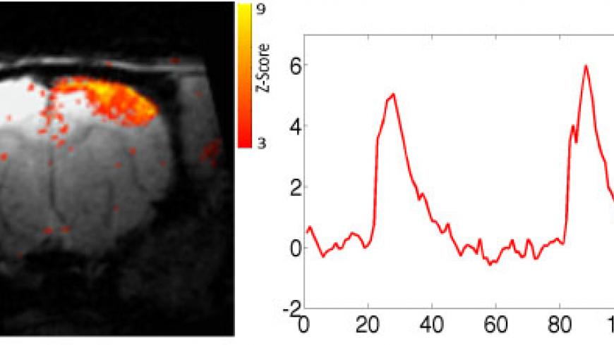
Functional MRI Z-maps (p<0.05) demonstrate increased BOLD responses in primary somatosensory cortex contralateral to forepaw stimulation in rats. Resolution: 100 × 100 × 1000 microns
(Image courtesy of Dr. Galit Pelled )
Diffusion Tensor Imaging (DTI)
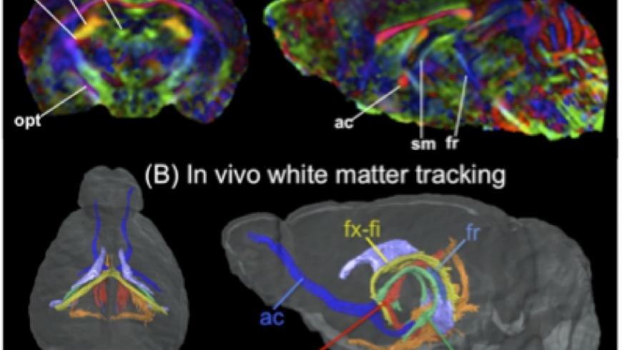
Comparison of 3D in vivo and ex vivo DTI images of adult mouse brains. The in vivo data (A) were linearly registered to ex vivo data. (B) White matter tracts reconstructed from the in vivo DTI data. Abbreviations: ac - anterior commissure; opt - optical track; fx-fi - fornix-fimbria; sm - stria medullaris; st - stria terminalis; fr - fasciculus retroflexus.
(Image courtesy of Dr. Jiangyang Zhang and Dr. Susumu Mori)
High Resolution Neuroanatomy
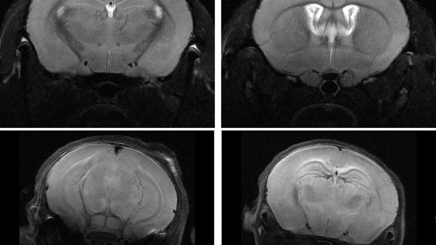
T2 weighted images of adult mouse and P15 mouse record using RARE sequence on Cyroprobe.
(Image courtesy of Dr. Jiangyang Zhang)
Cardiac MR Imaging
Triggered FLASH cine SAX movie of the healthy mouse heart.
(Image courtesy of Dr. Robert Weiss and Dr. Siamak Ardekani)