
Bruker Biospec 117/16 Ultra Shielded Refrigerated (USR) system is the first horizontal MRI system at 11.7 Tesla field strength. It delivers unsurpassed signal-to-noise ratio and resolution comparing to lower field small animal MRI systems. The bore size of the magnet is 155 mm, which provide easy access for imaging of rats and mouse. The drift rate for this magnet is 0.05 ppm/hr, and the homogeneity of the field is < 2 ppm for dsv 30 mm. The magnet is equipped with a two-stage Cryo-refrigerator (9 kW) which eliminates the need for liquid nitrogen and prolongs the liquid helium holding time. A Motorised Animal Positioning System is available on this scanner, designed to simplify animal handling and to increase animal throughput. Surface coils and head coils that are mounted onto the animal bed are also available with the system. Water beds are integrated in the animal beds to maintain body temperature during anesthesia. Laser crosshairs provide precise positioning of the region of interest. Repositioning accuracy is better than 1 mm.
The new integrated shim and actively shielded gradient system, B-GA 9S, provides extra high-order shim control Z3 and Z4.
Gradient specifications:
- Outer/inner diameter: 152 mm / 90 mm
- Gradient strength: 740 mT/m
- x, y, and z-linearity 60 mm dsv: < 5%
- Linear inductive rise time (x,y,z): 100 micro-second
- Slew rate: 6,600 T/m/s
The AVANCE III console provides highest speed RF generation and data acquisition with a highly modular and scalable transmitter and receiver channel architecture. Eight transmitters and eight receivers are available for this MRI scanner. Each receiver provides gating and power for the use of an external preamplifier. The system contains two 1H preamplifiers, one for high power and one for low power RF thresholds, and one preamplifier for X-nuclei.
Softwares
- The ParaVision 5.1 software package enables acquisition and reconstruction of data sets employing standard MRI techniques as well as user-defined MRI schemes.
- The ParaVision Spectroscopy Package consisting of TOPSPIN 2.0 for NMR data acquisition, processing, plotting, analysis and simulation as well as MRS techniques, such as localized single voxel spectroscopy and Spectroscopic imaging (CSI).
- DTI Evaluation Tools can perform calculation of tensor components and scalar invariants (tensor trace, anisotropy index); The DTI Visualization Tools is able to display maps of tensor components , scalar invariants and diffusion direction.
The papers published by investigators are based on data obtained using the Bruker Biospec 11.7T system (1-176).
A special thanks to Dr. Jiadi Xu and Dr. Yuguo Li for compiling the list.
1. Airan RD, Bar-Shir A, Liu G, Pelled G, McMahon MT, van Zijl PC et al. MRI biosensor for protein kinase A encoded by a single synthetic gene. Magn Reson Med 2012;68(6):1919-1923.
2. Chan KW, McMahon MT, Kato Y, Liu G, Bulte JW, Bhujwalla ZM et al. Natural D-glucose as a biodegradable MRI contrast agent for detecting cancer. Magn Reson Med 2012;68(6):1764-1773.
3. Gorelik M, Orukari I, Wang J, Galpoththawela S, Kim H, Levy M et al. Use of MR cell tracking to evaluate targeting of glial precursor cells to inflammatory tissue by exploiting the very late antigen-4 docking receptor. Radiology 2012;265(1):175-185.
4. Kim H, Walczak P, Kerr C, Galpoththawela C, Gilad AA, Muja N et al. Immunomodulation by transplanted human embryonic stem cell-derived oligodendroglial progenitors in experimental autoimmune encephalomyelitis. Stem Cells 2012;30(12):2820-2829.
5. Kim H, Walczak P, Muja N, Campanelli JT, Bulte JW. ICV-transplanted human glial precursor cells are short-lived yet exert immunomodulatory effects in mice with EAE. Glia 2012;60(7):1117-1129.
6. Liu G, Moake M, Har-el YE, Long CM, Chan KW, Cardona A et al. In vivo multicolor molecular MR imaging using diamagnetic chemical exchange saturation transfer liposomes. Magn Reson Med 2012;67(4):1106-1113.
7. Song X, Gilad AA, Joel S, Liu G, Bar-Shir A, Liang Y et al. CEST phase mapping using a length and offset varied saturation (LOVARS) scheme. Magn Reson Med 2012;68(4):1074-1086.
8. Aggarwal M, Zhang J, Pletnikova O, Crain B, Troncoso J, Mori S. Feasibility of creating a high-resolution 3D diffusion tensor imaging based atlas of the human brainstem: a case study at 11.7 T. Neuroimage 2013;74:117-127.
9. Bar-Shir A, Gilad AA, Chan KW, Liu G, van Zijl PC, Bulte JW et al. Metal ion sensing using ion chemical exchange saturation transfer 19F magnetic resonance imaging. J Am Chem Soc 2013;135(33):12164-12167.
10. Bar-Shir A, Liu G, Greenberg MM, Bulte JW, Gilad AA. Synthesis of a probe for monitoring HSV1-tk reporter gene expression using chemical exchange saturation transfer MRI. Nat Protoc 2013;8(12):2380-2391.
11. Bar-Shir A, Liu G, Liang Y, Yadav NN, McMahon MT, Walczak P et al. Transforming thymidine into a magnetic resonance imaging probe for monitoring gene expression. J Am Chem Soc 2013;135(4):1617-1624.
12. Barczewska M, Wojtkiewicz J, Habich A, Janowski M, Adamiak Z, Holak P et al. MR monitoring of minimally invasive delivery of mesenchymal stem cells into the porcine intervertebral disc. PLoS One 2013;8(9):e74658.
13. Chan KW, Liu G, Song X, Kim H, Yu T, Arifin DR et al. MRI-detectable pH nanosensors incorporated into hydrogels for in vivo sensing of transplanted-cell viability. Nat Mater 2013;12(3):268-275.
14. Cromer Berman SM, Kshitiz, Wang CJ, Orukari I, Levchenko A, Bulte JW et al. Cell motility of neural stem cells is reduced after SPIO-labeling, which is mitigated after exocytosis. Magn Reson Med 2013;69(1):255-262.
15. Han Y, Li N, Zeiler SR, Pelled G. Peripheral nerve injury induces immediate increases in layer v neuronal activity. Neurorehabil Neural Repair 2013;27(7):664-672.
16. Janowski M, Lyczek A, Engels C, Xu J, Lukomska B, Bulte JW et al. Cell size and velocity of injection are major determinants of the safety of intracarotid stem cell transplantation. J Cereb Blood Flow Metab 2013;33(6):921-927.
17. Liu G, Bettegowda C, Qiao Y, Staedtke V, Chan KW, Bai R et al. Noninvasive imaging of infection after treatment with tumor-homing bacteria using Chemical Exchange Saturation Transfer (CEST) MRI. Magn Reson Med 2013;70(6):1690-1698.
18. Liu G, Chan KW, Song X, Zhang J, Gilad AA, Bulte JW et al. NOrmalized MAgnetization Ratio (NOMAR) filtering for creation of tissue selective contrast maps. Magn Reson Med 2013;69(2):516-523.
19. Wu D, Xu J, McMahon MT, van Zijl PC, Mori S, Northington FJ et al. In vivo high-resolution diffusion tensor imaging of the mouse brain. Neuroimage 2013;83:18-26.
20. Yadav NN, Chan KW, Jones CK, McMahon MT, van Zijl PC. Time domain removal of irrelevant magnetization in chemical exchange saturation transfer Z-spectra. Magn Reson Med 2013;70(2):547-555.
21. Yang X, Song X, Li Y, Liu G, Banerjee SR, Pomper MG et al. Salicylic acid and analogues as diaCEST MRI contrast agents with highly shifted exchangeable proton frequencies. Angew Chem Int Ed Engl 2013;52(31):8116-8119.
22. Bar-Shir A, Liu G, Chan KW, Oskolkov N, Song X, Yadav NN et al. Human protamine-1 as an MRI reporter gene based on chemical exchange. ACS Chem Biol 2014;9(1):134-138.
23. Chan KW, Bulte JW, McMahon MT. Diamagnetic chemical exchange saturation transfer (diaCEST) liposomes: physicochemical properties and imaging applications. Wiley Interdiscip Rev Nanomed Nanobiotechnol 2014;6(1):111-124.
24. Chan KW, Liu G, van Zijl PC, Bulte JW, McMahon MT. Magnetization transfer contrast MRI for non-invasive assessment of innate and adaptive immune responses against alginate-encapsulated cells. Biomaterials 2014;35(27):7811-7818.
25. Chan KW, Yu T, Qiao Y, Liu Q, Yang M, Patel H et al. A diaCEST MRI approach for monitoring liposomal accumulation in tumors. J Control Release 2014;180:51-59.
26. Dada T, Rosenzweig JM, Al Shammary M, Firdaus W, Al Rebh S, Borbiev T et al. Mouse model of intrauterine inflammation: sex-specific differences in long-term neurologic and immune sequelae. Brain Behav Immun 2014;38:142-150.
27. Janowski M, Engels C, Gorelik M, Lyczek A, Bernard S, Bulte JW et al. Survival of neural progenitors allografted into the CNS of immunocompetent recipients is highly dependent on transplantation site. Cell Transplant 2014;23(2):253-262.
28. Li N, Yang Y, Glover DP, Zhang J, Saraswati M, Robertson C et al. Evidence for impaired plasticity after traumatic brain injury in the developing brain. J Neurotrauma 2014;31(4):395-403.
29. Liu G, Qin Q, Chan KW, Li Y, Bulte JW, McMahon MT et al. Non-invasive temperature mapping using temperature-responsive water saturation shift referencing (T-WASSR) MRI. NMR Biomed 2014;27(3):320-331.
30. Wu D, Martin LJ, Northington FJ, Zhang J. Oscillating gradient diffusion MRI reveals unique microstructural information in normal and hypoxia-ischemia injured mouse brains. Magn Reson Med 2014;72(5):1366-1374.
31. Wu D, Reisinger D, Xu J, Fatemi SA, van Zijl PC, Mori S et al. Localized diffusion magnetic resonance micro-imaging of the live mouse brain. Neuroimage 2014;91:12-20.
32. Xu J, Yadav NN, Bar-Shir A, Jones CK, Chan KW, Zhang J et al. Variable delay multi-pulse train for fast chemical exchange saturation transfer and relayed-nuclear overhauser enhancement MRI. Magn Reson Med 2014;71(5):1798-1812.
33. Yadav NN, Xu J, Bar-Shir A, Qin Q, Chan KW, Grgac K et al. Natural D-glucose as a biodegradable MRI relaxation agent. Magn Reson Med 2014;72(3):823-828.
34. Bar-Shir A, Bulte JW, Gilad AA. Molecular engineering of nonmetallic biosensors for CEST MRI. ACS Chem Biol 2015;10(5):1160-1170.
35. Bar-Shir A, Liang Y, Chan KW, Gilad AA, Bulte JW. Supercharged green fluorescent proteins as bimodal reporter genes for CEST MRI and optical imaging. Chem Commun (Camb) 2015;51(23):4869-4871.
36. Bar-Shir A, Yadav NN, Gilad AA, van Zijl PC, McMahon MT, Bulte JW. Single (19)F probe for simultaneous detection of multiple metal ions using miCEST MRI. J Am Chem Soc 2015;137(1):78-81.
37. Burnsed JC, Chavez-Valdez R, Hossain MS, Kesavan K, Martin LJ, Zhang J et al. Hypoxia-ischemia and therapeutic hypothermia in the neonatal mouse brain--a longitudinal study. PLoS One 2015;10(3):e0118889.
38. Farrar CT, Buhrman JS, Liu G, Kleijn A, Lamfers ML, McMahon MT et al. Establishing the Lysine-rich Protein CEST Reporter Gene as a CEST MR Imaging Detector for Oncolytic Virotherapy. Radiology 2015;275(3):746-754.
39. Kadayakkara DK, Korrer MJ, Bulte JW, Levitsky HI. Paradoxical decrease in the capture and lymph node delivery of cancer vaccine antigen induced by a TLR4 agonist as visualized by dual-mode imaging. Cancer Res 2015;75(1):51-61.
40. Liang Y, Bar-Shir A, Song X, Gilad AA, Walczak P, Bulte JW. Label-free imaging of gelatin-containing hydrogel scaffolds. Biomaterials 2015;42:144-150.
41. Lu H, Kobilo T, Robertson C, Tong S, Celnik P, Pelled G. Transcranial magnetic stimulation facilitates neurorehabilitation after pediatric traumatic brain injury. Sci Rep 2015;5:14769.
42. Minn I, Bar-Shir A, Yarlagadda K, Bulte JW, Fisher PB, Wang H et al. Tumor-specific expression and detection of a CEST reporter gene. Magn Reson Med 2015;74(2):544-549.
43. Oskolkov N, Bar-Shir A, Chan KW, Song X, van Zijl PC, Bulte JW et al. Biophysical Characterization of Human Protamine-1 as a Responsive CEST MR Contrast Agent. ACS Macro Lett 2015;4(1):34-38.
44. Schunke KJ, Toung TK, Zhang J, Pathak AP, Xu J, Zhang J et al. A novel atherothrombotic model of ischemic stroke induced by injection of collagen into the cerebral vasculature. J Neurosci Methods 2015;239:65-74.
45. Song X, Airan RD, Arifin DR, Bar-Shir A, Kadayakkara DK, Liu G et al. Label-free in vivo molecular imaging of underglycosylated mucin-1 expression in tumour cells. Nat Commun 2015;6:6719.
46. Song X, Xu J, Xia S, Yadav NN, Lal B, Laterra J et al. Multi-echo length and offset VARied saturation (MeLOVARS) method for improved CEST imaging. Magn Reson Med 2015;73(2):488-496.
47. Song X, Yang X, Ray Banerjee S, Pomper MG, McMahon MT. Anthranilic acid analogs as diamagnetic CEST MRI contrast agents that feature an intramolecular-bond shifted hydrogen. Contrast Media Mol Imaging 2015;10(1):74-80.
48. Thatipamula S, Al Rahim M, Zhang J, Hossain MA. Genetic deletion of neuronal pentraxin 1 expression prevents brain injury in a neonatal mouse model of cerebral hypoxia-ischemia. Neurobiol Dis 2015;75:15-30.
49. Wu D, Lei J, Rosenzweig JM, Burd I, Zhang J. In utero localized diffusion MRI of the embryonic mouse brain microstructure and injury. J Magn Reson Imaging 2015;42(3):717-728.
50. Xu X, Chan KW, Knutsson L, Artemov D, Xu J, Liu G et al. Dynamic glucose enhanced (DGE) MRI for combined imaging of blood-brain barrier break down and increased blood volume in brain cancer. Magn Reson Med 2015;74(6):1556-1563.
51. Yu T, Chan KW, Anonuevo A, Song X, Schuster BS, Chattopadhyay S et al. Liposome-based mucus-penetrating particles (MPP) for mucosal theranostics: demonstration of diamagnetic chemical exchange saturation transfer (diaCEST) magnetic resonance imaging (MRI). Nanomedicine 2015;11(2):401-405.
52. Aggarwal M, Kageyama Y, Li X, van Zijl PC. B0 -orientation dependent magnetic susceptibility-induced white matter contrast in the human brainstem at 11.7T. Magn Reson Med 2016;75(6):2455-2463.
53. Chan KW, Jiang L, Cheng M, Wijnen JP, Liu G, Huang P et al. CEST-MRI detects metabolite levels altered by breast cancer cell aggressiveness and chemotherapy response. NMR Biomed 2016;29(6):806-816.
54. Chehade M, Srivastava AK, Bulte JW. Co-Registration of Bioluminescence Tomography, Computed Tomography, and Magnetic Resonance Imaging for Multimodal In Vivo Stem Cell Tracking. Tomography 2016;2(2):159-165.
55. Jusue-Torres I, Jeon LH, Sankey EW, Lu J, Vivas-Buitrago T, Crawford JA et al. A Novel Experimental Animal Model of Adult Chronic Hydrocephalus. Neurosurgery 2016;79(5):746-756.
56. Lesniak WG, Oskolkov N, Song X, Lal B, Yang X, Pomper M et al. Salicylic Acid Conjugated Dendrimers Are a Tunable, High Performance CEST MRI NanoPlatform. Nano Lett 2016;16(4):2248-2253.
57. Li W, Grgac K, Huang A, Yadav N, Qin Q, van Zijl PC. Quantitative theory for the longitudinal relaxation time of blood water. Magn Reson Med 2016;76(1):270-281.
58. Li Y, Chen H, Xu J, Yadav NN, Chan KW, Luo L et al. CEST theranostics: label-free MR imaging of anticancer drugs. Oncotarget 2016;7(6):6369-6378.
59. Liu H, Jablonska A, Li Y, Cao S, Liu D, Chen H et al. Label-free CEST MRI Detection of Citicoline-Liposome Drug Delivery in Ischemic Stroke. Theranostics 2016;6(10):1588-1600.
60. Ngen EJ, Bar-Shir A, Jablonska A, Liu G, Song X, Ansari R et al. Imaging the DNA Alkylator Melphalan by CEST MRI: An Advanced Approach to Theranostics. Mol Pharm 2016;13(9):3043-3053.
61. Song X, Walczak P, He X, Yang X, Pearl M, Bulte JWM et al. Salicylic acid analogues as chemical exchange saturation transfer MRI contrast agents for the assessment of brain perfusion territory and blood-brain barrier opening after intra-arterial infusion. J Cereb Blood Flow Metab 2016;36(7):1186-1194.
62. Wu D, Zhang J. In vivo mapping of macroscopic neuronal projections in the mouse hippocampus using high-resolution diffusion MRI. Neuroimage 2016;125:84-93.
63. Xu J, Qin Q, Wu D, Hua J, Song X, McMahon MT et al. Steady pulsed imaging and labeling scheme for noninvasive perfusion imaging. Magn Reson Med 2016;75(1):238-248.
64. Xu X, Yadav NN, Song X, McMahon MT, Jerschow A, van Zijl PC et al. Screening CEST contrast agents using ultrafast CEST imaging. J Magn Reson 2016;265:224-229.
65. Xu X, Yadav NN, Zeng H, Jones CK, Zhou J, van Zijl PC et al. Magnetization transfer contrast-suppressed imaging of amide proton transfer and relayed nuclear overhauser enhancement chemical exchange saturation transfer effects in the human brain at 7T. Magn Reson Med 2016;75(1):88-96.
66. Yang X, Song X, Ray Banerjee S, Li Y, Byun Y, Liu G et al. Developing imidazoles as CEST MRI pH sensors. Contrast Media Mol Imaging 2016;11(4):304-312.
67. Airan RD, Meyer RA, Ellens NP, Rhodes KR, Farahani K, Pomper MG et al. Noninvasive Targeted Transcranial Neuromodulation via Focused Ultrasound Gated Drug Release from Nanoemulsions. Nano Lett 2017;17(2):652-659.
68. Chen L, Zeng H, Xu X, Yadav NN, Cai S, Puts NA et al. Investigation of the contribution of total creatine to the CEST Z-spectrum of brain using a knockout mouse model. NMR Biomed 2017;30(12).
69. Grgac K, Li W, Huang A, Qin Q, van Zijl PC. Transverse water relaxation in whole blood and erythrocytes at 3T, 7T, 9.4T, 11.7T and 16.4T; determination of intracellular hemoglobin and extracellular albumin relaxivities. Magn Reson Imaging 2017;38:234-249.
70. Li Q, Li G, Wu D, Lu H, Hou Z, Ross CA et al. Resting-state functional MRI reveals altered brain connectivity and its correlation with motor dysfunction in a mouse model of Huntington's disease. Sci Rep 2017;7(1):16742.
71. Liu G, Banerjee SR, Yang X, Yadav N, Lisok A, Jablonska A et al. A dextran-based probe for the targeted magnetic resonance imaging of tumours expressing prostate-specific membrane antigen. Nat Biomed Eng 2017;1:977-982.
72. Lock LL, Li Y, Mao X, Chen H, Staedtke V, Bai R et al. One-Component Supramolecular Filament Hydrogels as Theranostic Label-Free Magnetic Resonance Imaging Agents. ACS Nano 2017;11(1):797-805.
73. Lyczek A, Arnold A, Zhang J, Campanelli JT, Janowski M, Bulte JW et al. Transplanted human glial-restricted progenitors can rescue the survival of dysmyelinated mice independent of the production of mature, compact myelin. Exp Neurol 2017;291:74-86.
74. Muhammad G, Xu J, Bulte JWM, Jablonska A, Walczak P, Janowski M. Transplanted adipose-derived stem cells can be short-lived yet accelerate healing of acid-burn skin wounds: a multimodal imaging study. Sci Rep 2017;7(1):4644.
75. Ryoo D, Xu X, Li Y, Tang JA, Zhang J, van Zijl PCM et al. Detection and Quantification of Hydrogen Peroxide in Aqueous Solutions Using Chemical Exchange Saturation Transfer. Anal Chem 2017;89(14):7758-7764.
76. Semenkow S, Li S, Kahlert UD, Raabe EH, Xu J, Arnold A et al. An immunocompetent mouse model of human glioblastoma. Oncotarget 2017;8(37):61072-61082.
77. Shin SH, Kadayakkara DK, Bulte JW. In Vivo (19)F MR Imaging Cell Tracking of Inflammatory Macrophages and Site-specific Development of Colitis-associated Dysplasia. Radiology 2017;282(1):194-201.
78. Walczak P, Wojtkiewicz J, Nowakowski A, Habich A, Holak P, Xu J et al. Real-time MRI for precise and predictable intra-arterial stem cell delivery to the central nervous system. J Cereb Blood Flow Metab 2017;37(7):2346-2358.
79. Wu D, Zhang J. The Effect of Microcirculatory Flow on Oscillating Gradient Diffusion MRI and Diffusion Encoding with Dual-Frequency Orthogonal Gradients (DEFOG). Magn Reson Med 2017;77(4):1583-1592.
80. Xu J, Chan KW, Xu X, Yadav N, Liu G, van Zijl PC. On-resonance variable delay multipulse scheme for imaging of fast-exchanging protons and semisolid macromolecules. Magn Reson Med 2017;77(2):730-739.
81. Yadav NN, Yang X, Li Y, Li W, Liu G, van Zijl PCM. Detection of dynamic substrate binding using MRI. Sci Rep 2017;7(1):10138.
82. Yang J, Li Q, Wang Z, Qi C, Han X, Lan X et al. Multimodality MRI assessment of grey and white matter injury and blood-brain barrier disruption after intracerebral haemorrhage in mice. Sci Rep 2017;7:40358.
83. Aggarwal M, Li X, Grohn O, Sierra A. Nuclei-specific deposits of iron and calcium in the rat thalamus after status epilepticus revealed with quantitative susceptibility mapping (QSM). J Magn Reson Imaging 2018;47(2):554-564.
84. Banerjee SR, Song X, Yang X, Minn I, Lisok A, Chen Y et al. Salicylic Acid-Based Polymeric Contrast Agents for Molecular Magnetic Resonance Imaging of Prostate Cancer. Chemistry 2018;24(28):7235-7242.
85. Bar-Shir A, Alon L, Korrer MJ, Lim HS, Yadav NN, Kato Y et al. Quantification and tracking of genetically engineered dendritic cells for studying immunotherapy. Magn Reson Med 2018;79(2):1010-1019.
86. Chen L, Xu X, Zeng H, Chan KWY, Yadav N, Cai S et al. Separating fast and slow exchange transfer and magnetization transfer using off-resonance variable-delay multiple-pulse (VDMP) MRI. Magn Reson Med 2018;80(4):1568-1576.
87. Chu C, Liu G, Janowski M, Bulte JWM, Li S, Pearl M et al. Real-Time MRI Guidance for Reproducible Hyperosmolar Opening of the Blood-Brain Barrier in Mice. Front Neurol 2018;9:921.
88. Doman SE, Girish A, Nemeth CL, Drummond GT, Carr P, Garcia MS et al. Early Detection of Hypothermic Neuroprotection Using T2-Weighted Magnetic Resonance Imaging in a Mouse Model of Hypoxic Ischemic Encephalopathy. Front Neurol 2018;9:304.
89. Li Y, Qiao Y, Chen H, Bai R, Staedtke V, Han Z et al. Characterization of tumor vascular permeability using natural dextrans and CEST MRI. Magn Reson Med 2018;79(2):1001-1009.
90. Liang Y, Zhang J, Walczak P, Bulte JWM. Quantification of motor neuron loss and muscular atrophy in ricin-induced focal nerve injury. J Neurosci Methods 2018;308:142-150.
91. Liu J, Bai R, Li Y, Staedtke V, Zhang S, van Zijl PCM et al. MRI detection of bacterial brain abscesses and monitoring of antibiotic treatment using bacCEST. Magn Reson Med 2018;80(2):662-671.
92. Liu J, Han Z, Chen G, Li Y, Zhang J, Xu J et al. CEST MRI of sepsis-induced acute kidney injury. NMR Biomed 2018;31(8):e3942.
93. Vivas-Buitrago T, Pinilla-Monsalve G, Jusue-Torres I, Oishi K, Robison J, Crawford JA et al. Ventricular Volume Dynamics During the Development of Adult Chronic Communicating Hydrocephalus in a Rodent Model. World Neurosurg 2018;120:e1120-e1127.
94. Wei Z, Xu J, Liu P, Chen L, Li W, van Zijl P et al. Quantitative assessment of cerebral venous blood T(2) in mouse at 11.7T: Implementation, optimization, and age effect. Magn Reson Med 2018;80(2):521-528.
95. Wu D, Lei J, Jia B, Xie H, Zhu Y, Xu J et al. In vivo assessment of the placental anatomy and perfusion in a mouse model of intrauterine inflammation. J Magn Reson Imaging 2018;47(5):1260-1267.
96. Wu D, Li Q, Northington FJ, Zhang J. Oscillating gradient diffusion kurtosis imaging of normal and injured mouse brains. NMR Biomed 2018;31(6):e3917.
97. Wu D, Xu J, Lei J, McLane M, van Zijl PC, Burd I. Dynamic glucose enhanced MRI of the placenta in a mouse model of intrauterine inflammation. Placenta 2018;69:86-91.
98. Zhang J, Wu D, Turnbull DH. In Utero MRI of Mouse Embryos. Methods Mol Biol 2018;1718:285-296.
99. Arifin DR, Kulkarni M, Kadayakkara D, Bulte JWM. Fluorocapsules allow in vivo monitoring of the mechanical stability of encapsulated islet cell transplants. Biomaterials 2019;221:119410.
100. Breu M, Reisinger D, Tao L, Wu D, Zhang Y, Budde MD et al. In vivo high-resolution diffusion tensor imaging of the developing neonatal rat cortex and its relationship to glial and dendritic maturation. Brain Struct Funct 2019;224(5):1815-1829.
101. Chen H, Liu D, Li Y, Xu X, Xu J, Yadav NN et al. CEST MRI monitoring of tumor response to vascular disrupting therapy using high molecular weight dextrans. Magn Reson Med 2019;82(4):1471-1479.
102. Chen L, Barker PB, Weiss RG, van Zijl PCM, Xu J. Creatine and phosphocreatine mapping of mouse skeletal muscle by a polynomial and Lorentzian line-shape fitting CEST method. Magn Reson Med 2019;81(1):69-78.
103. Chen L, Wei Z, Cai S, Li Y, Liu G, Lu H et al. High-resolution creatine mapping of mouse brain at 11.7 T using non-steady-state chemical exchange saturation transfer. NMR Biomed 2019;32(11):e4168.
104. Chen L, Wei Z, Chan KWY, Cai S, Liu G, Lu H et al. Protein aggregation linked to Alzheimer's disease revealed by saturation transfer MRI. Neuroimage 2019;188:380-390.
105. Chen Z, Li Y, Airan R, Han Z, Xu J, Chan KWY et al. CT and CEST MRI bimodal imaging of the intratumoral distribution of iodinated liposomes. Quant Imaging Med Surg 2019;9(9):1579-1591.
106. Eloundou SN, Lee J, Wu D, Lei J, Feller MC, Ozen M et al. Placental malperfusion in response to intrauterine inflammation and its connection to fetal sequelae. PLoS One 2019;14(4):e0214951.
107. Gullotti D, Mangraviti A, Zhang J, Volpin F, Fink G, Reiss R et al. Evaluation of select biocompatible markers for labelling peripheral nerves on 11.7 T MRI. J Neurosci Methods 2019;315:6-13.
108. Han Z, Li Y, Zhang J, Liu J, Chen C, van Zijl PC et al. Molecular Imaging of Deoxycytidine Kinase Activity Using Deoxycytidine-Enhanced CEST MRI. Cancer Res 2019;79(10):2775-2783.
109. Han Z, Zhang S, Fujiwara K, Zhang J, Li Y, Liu J et al. Extradomain-B Fibronectin-Targeted Dextran-Based Chemical Exchange Saturation Transfer Magnetic Resonance Imaging Probe for Detecting Pancreatic Cancer. Bioconjug Chem 2019;30(5):1425-1433.
110. Krishnan VS, Shin SS, Belegu V, Celnik P, Reimers M, Smith KR et al. Multimodal Evaluation of TMS - Induced Somatosensory Plasticity and Behavioral Recovery in Rats With Contusion Spinal Cord Injury. Front Neurosci 2019;13:387.
111. Liu T, Chen Y, Thomas AM, Song X. CEST MRI with distribution-based analysis for assessment of early stage disease activity in a mouse model of multiple sclerosis: An initial study. NMR Biomed 2019;32(11):e4139.
112. Ngen EJ, Benham Azad B, Boinapally S, Lisok A, Brummet M, Jacob D et al. MRI Assessment of Prostate-Specific Membrane Antigen (PSMA) Targeting by a PSMA-Targeted Magnetic Nanoparticle: Potential for Image-Guided Therapy. Mol Pharm 2019;16(5):2060-2068.
113. Pavuluri K, Manoli I, Pass A, Li Y, Vernon HJ, Venditti CP et al. Noninvasive monitoring of chronic kidney disease using pH and perfusion imaging. Sci Adv 2019;5(8):eaaw8357.
114. Richard JP, Hussain U, Gross S, Taga A, Kouser M, Almad A et al. Perfluorocarbon Labeling of Human Glial-Restricted Progenitors for (19) F Magnetic Resonance Imaging. Stem Cells Transl Med 2019;8(4):355-365.
115. Sehgal AA, Li Y, Lal B, Yadav NN, Xu X, Xu J et al. CEST MRI of 3-O-methyl-D-glucose uptake and accumulation in brain tumors. Magn Reson Med 2019;81(3):1993-2000.
116. Srivastava RK, Jablonska A, Chu C, Gregg L, Bulte JWM, Koehler RC et al. Biodistribution of Glial Progenitors in a Three Dimensional-Printed Model of the Piglet Cerebral Ventricular System. Stem Cells Dev 2019;28(8):515-527.
117. Vivas-Buitrago T, Lokossou A, Jusue-Torres I, Pinilla-Monsalve G, Blitz AM, Herzka DA et al. Aqueductal Cerebrospinal Fluid Stroke Volume Flow in a Rodent Model of Chronic Communicating Hydrocephalus: Establishing a Homogeneous Study Population for Cerebrospinal Fluid Dynamics Exploration. World Neurosurg 2019;128:e1118-e1125.
118. Wei Z, Chen L, Lin Z, Jiang D, Xu J, Liu P et al. Optimization of phase-contrast MRI for the estimation of global cerebral blood flow of mice at 11.7T. Magn Reson Med 2019;81(4):2566-2575.
119. Wu D, Martin LJ, Northington FJ, Zhang J. Oscillating-gradient diffusion magnetic resonance imaging detects acute subcellular structural changes in the mouse forebrain after neonatal hypoxia-ischemia. J Cereb Blood Flow Metab 2019;39(7):1336-1348.
120. Xu X, Xu J, Chan KWY, Liu J, Liu H, Li Y et al. GlucoCEST imaging with on-resonance variable delay multiple pulse (onVDMP) MRI. Magn Reson Med 2019;81(1):47-56.
121. Xu X, Xu J, Knutsson L, Liu J, Liu H, Li Y et al. The effect of the mTOR inhibitor rapamycin on glucoCEST signal in a preclinical model of glioblastoma. Magn Reson Med 2019;81(6):3798-3807.
122. Yuan Y, Wei Z, Chu C, Zhang J, Song X, Walczak P et al. Development of Zinc-Specific iCEST MRI as an Imaging Biomarker for Prostate Cancer. Angew Chem Int Ed Engl 2019;58(43):15512-15517.
123. Yuan Y, Zhang J, Qi X, Li S, Liu G, Siddhanta S et al. Furin-mediated intracellular self-assembly of olsalazine nanoparticles for enhanced magnetic resonance imaging and tumour therapy. Nat Mater 2019;18(12):1376-1383.
124. Zhang J, Yuan Y, Gao M, Han Z, Chu C, Li Y et al. Carbon Dots as a New Class of Diamagnetic Chemical Exchange Saturation Transfer (diaCEST) MRI Contrast Agents. Angew Chem Int Ed Engl 2019;58(29):9871-9875.
125. Zhang J, Yuan Y, Han Z, Li Y, van Zijl PCM, Yang X et al. Detecting acid phosphatase enzymatic activity with phenol as a chemical exchange saturation transfer magnetic resonance imaging contrast agent (PhenolCEST MRI). Biosens Bioelectron 2019;141:111442.
126. Zhu W, Chu C, Kuddannaya S, Yuan Y, Walczak P, Singh A et al. In Vivo Imaging of Composite Hydrogel Scaffold Degradation Using CEST MRI and Two-Color NIR Imaging. Adv Funct Mater 2019;29(36).
127. Chen L, Cao S, Koehler RC, van Zijl PCM, Xu J. High-sensitivity CEST mapping using a spatiotemporal correlation-enhanced method. Magn Reson Med 2020;84(6):3342-3350.
128. Chu C, Davis CM, Lan X, Hienz RD, Jablonska A, Thomas AM et al. Neuroinflammation After Stereotactic Radiosurgery-Induced Brain Tumor Disintegration Is Linked to Persistent Cognitive Decline in a Mouse Model of Metastatic Disease. Int J Radiat Oncol Biol Phys 2020;108(3):745-757.
129. Chu C, Jablonska A, Lesniak WG, Thomas AM, Lan X, Linville RM et al. Optimization of osmotic blood-brain barrier opening to enable intravital microscopy studies on drug delivery in mouse cortex. J Control Release 2020;317:312-321.
130. Krishnan V, Xu J, Mendoza AG, Koretsky A, Anderson SA, Pelled G. High-resolution MEMRI characterizes laminar specific ascending and descending spinal cord pathways in rats. J Neurosci Methods 2020;340:108748.
131. Li W, van Zijl PCM. Quantitative theory for the transverse relaxation time of blood water. NMR Biomed 2020;33(5):e4207.
132. Nemeth CL, Tomlinson SN, Rosen M, O'Brien BM, Larraza O, Jain M et al. Neuronal ablation of mt-AspRS in mice induces immune pathway activation prior to severe and progressive cortical and behavioral disruption. Exp Neurol 2020;326:113164.
133. Ning B, Guo G, Gu C, Xu J, Bibic A, He X et al. Mutant G2019S-LRRK2 Induces Abnormalities in Arteriolar Cerebral Blood Volume in Mouse Brains: An MRI Study. Neurodegener Dis 2020;20(2-3):65-72.
134. Oishi K, Mori S, Troncoso JC, Lenz FA. Mapping tracts in the human subthalamic area by 11.7T ex vivo diffusion tensor imaging. Brain Struct Funct 2020;225(4):1293-1312.
135. Pavuluri K, Rosenberg JT, Helsper S, Bo S, McMahon MT. Amplified detection of phosphocreatine and creatine after supplementation using CEST MRI at high and ultrahigh magnetic fields. J Magn Reson 2020;313:106703.
136. Thomas AM, Li S, Chu C, Shats I, Xu J, Calabresi PA et al. Evaluation of cell transplant-mediated attenuation of diffuse injury in experimental autoimmune encephalomyelitis using onVDMP CEST MRI. Exp Neurol 2020;329:113316.
137. Thomas AM, Xu J, Calabresi PA, van Zijl PCM, Bulte JWM. Monitoring diffuse injury during disease progression in experimental autoimmune encephalomyelitis with on resonance variable delay multiple pulse (onVDMP) CEST MRI. Neuroimage 2020;204:116245.
138. Wei Z, Chen L, Hou X, van Zijl PCM, Xu J, Lu H. Age-Related Alterations in Brain Perfusion, Venous Oxygenation, and Oxygen Metabolic Rate of Mice: A 17-Month Longitudinal MRI Study. Front Neurol 2020;11:559.
139. Wei Z, Wang Q, Modi HR, Cho SM, Geocadin R, Thakor NV et al. Acute-stage MRI cerebral oxygen consumption biomarkers predict 24-hour neurological outcome in a rat cardiac arrest model. NMR Biomed 2020;33(11):e4377.
140. Wu D, Lei J, Xie H, Dong J, Burd I. Diffusion MRI revealed altered inter-hippocampal projections in the mouse brain after intrauterine inflammation. Brain Imaging Behav 2020;14(2):383-395.
141. Zhou Y, van Zijl PCM, Xu X, Xu J, Li Y, Chen L et al. Magnetic resonance imaging of glycogen using its magnetic coupling with water. Proc Natl Acad Sci U S A 2020;117(6):3144-3149.
142. Bhargava P, Kim S, Reyes AA, Grenningloh R, Boschert U, Absinta M et al. Imaging meningeal inflammation in CNS autoimmunity identifies a therapeutic role for BTK inhibition. Brain 2021;144(5):1396-1408.
143. Chary K, Narvaez O, Salo RA, San Martin Molina I, Tohka J, Aggarwal M et al. Microstructural Tissue Changes in a Rat Model of Mild Traumatic Brain Injury. Front Neurosci 2021;15:746214.
144. Chen L, Liu J, Chu C, Han Z, Yadav N, Xu J et al. Deuterium oxide as a contrast medium for real-time MRI-guided endovascular neurointervention. Theranostics 2021;11(13):6240-6250.
145. Chen L, van Zijl PCM, Wei Z, Lu H, Duan W, Wong PC et al. Early detection of Alzheimer's disease using creatine chemical exchange saturation transfer magnetic resonance imaging. Neuroimage 2021;236:118071.
146. Chen L, Wei Z, Chan KW, Li Y, Suchal K, Bi S et al. D-Glucose uptake and clearance in the tauopathy Alzheimer's disease mouse brain detected by on-resonance variable delay multiple pulse MRI. J Cereb Blood Flow Metab 2021;41(5):1013-1025.
147. Han Z, Liu S, Pei Y, Ding Z, Li Y, Wang X et al. Highly efficient magnetic labelling allows MRI tracking of the homing of stem cell-derived extracellular vesicles following systemic delivery. J Extracell Vesicles 2021;10(3):e12054.
148. Kuddannaya S, Zhu W, Chu C, Singh A, Walczak P, Bulte JWM. In Vivo Imaging of Allografted Glial-Restricted Progenitor Cell Survival and Hydrogel Scaffold Biodegradation. ACS Appl Mater Interfaces 2021;13(20):23423-23437.
149. Liu H, Zhang C, Xu J, Jin J, Cheng L, Miao X et al. Huntingtin silencing delays onset and slows progression of Huntington's disease: a biomarker study. Brain 2021;144(10):3101-3113.
150. Ngen EJ, Chen Y, Azad BB, Boinapally S, Jacob D, Lisok A et al. Prostate-specific membrane antigen (PSMA)-targeted photodynamic therapy enhances the delivery of PSMA-targeted magnetic nanoparticles to PSMA-expressing prostate tumors. Nanotheranostics 2021;5(2):182-196.
151. Wang R, Chu C, Wei Z, Chen L, Xu J, Liang Y et al. Traumatic brain injury does not disrupt costimulatory blockade-induced immunological tolerance to glial-restricted progenitor allografts. J Neuroinflammation 2021;18(1):104.
152. Wei Z, Xu J, Chen L, Hirschler L, Barbier EL, Li T et al. Brain metabolism in tau and amyloid mouse models of Alzheimer's disease: An MRI study. NMR Biomed 2021;34(9):e4568.
153. Zhou Y, van Zijl PCM, Xu J, Yadav NN. Mechanism and quantitative assessment of saturation transfer for water-based detection of the aliphatic protons in carbohydrate polymers. Magn Reson Med 2021;85(3):1643-1654.
154. Bie C, Li Y, Zhou Y, Bhujwalla ZM, Song X, Liu G et al. Deep learning-based classification of preclinical breast cancer tumor models using chemical exchange saturation transfer magnetic resonance imaging. NMR Biomed 2022;35(2):e4626.
155. Bo S, Stabinska J, Wu Y, Pavuluri KD, Singh A, Mohanta Z et al. Exploring the potential of the novel imidazole-4,5-dicarboxyamide chemical exchange saturation transfer scaffold for pH and perfusion imaging. NMR Biomed 2022:e4894.
156. Chu C, Jablonska A, Gao Y, Lan X, Lesniak WG, Liang Y et al. Hyperosmolar blood-brain barrier opening using intra-arterial injection of hyperosmotic mannitol in mice under real-time MRI guidance. Nat Protoc 2022;17(1):76-94.
157. Guo Y, Cho SM, Wei Z, Wang Q, Modi HR, Gharibani P et al. Early Thalamocortical Reperfusion Leads to Neurologic Recovery in a Rodent Cardiac Arrest Model. Neurocrit Care 2022;37(1):60-72.
158. Han Z, Chen C, Xu X, Bai R, Staedtke V, Huang J et al. Dynamic contrast-enhanced CEST MRI using a low molecular weight dextran. NMR Biomed 2022;35(3):e4649.
159. Knutsson L, Xu X, van Zijl PCM, Chan KWY. Imaging of sugar-based contrast agents using their hydroxyl proton exchange properties. NMR Biomed 2022:e4784.
160. Kuddannaya S, Zhu W, Bulte JWM. In Vivo Imaging of Implanted Hyaluronic Acid Hydrogel Biodegradation. Methods Mol Biol 2022;2394:743-765.
161. Li AM, Chen L, Liu H, Li Y, Duan W, Xu J. Age-dependent cerebrospinal fluid-tissue water exchange detected by magnetization transfer indirect spin labeling MRI. Magn Reson Med 2022;87(5):2287-2298.
162. Liu J, Chu C, Zhang J, Bie C, Chen L, Aafreen S et al. Label-Free Assessment of Mannitol Accumulation Following Osmotic Blood-Brain Barrier Opening Using Chemical Exchange Saturation Transfer Magnetic Resonance Imaging. Pharmaceutics 2022;14(11).
163. Lyden PD, Bosetti F, Diniz MA, Rogatko A, Koenig JI, Lamb J et al. The Stroke Preclinical Assessment Network: Rationale, Design, Feasibility, and Stage 1 Results. Stroke 2022;53(5):1802-1812.
164. Pavuluri K, Yang E, Ayyappan V, Sonkar K, Tan Z, Tressler CM et al. Unlabeled aspirin as an activatable theranostic MRI agent for breast cancer. Theranostics 2022;12(4):1937-1951.
165. Thomas AM, Yang E, Smith MD, Chu C, Calabresi PA, Glunde K et al. CEST MRI and MALDI imaging reveal metabolic alterations in the cervical lymph nodes of EAE mice. J Neuroinflammation 2022;19(1):130.
166. Wei Z, Li Y, Hou X, Han Z, Xu J, McMahon MT et al. Quantitative cerebrovascular reactivity MRI in mice using acetazolamide challenge. Magn Reson Med 2022;88(5):2233-2241.
167. Wu D, Richards LJ, Zhao Z, Cao Z, Luo W, Shao W et al. A diffusion MRI-based spatiotemporal continuum of the embryonic mouse brain for probing gene-neuroanatomy connections. Proc Natl Acad Sci U S A 2022;119(7).
168. Yuan Y, Wang C, Kuddannaya S, Zhang J, Arifin DR, Han Z et al. In vivo tracking of unlabelled mesenchymal stromal cells by mannose-weighted chemical exchange saturation transfer MRI. Nat Biomed Eng 2022;6(5):658-666.
169. Zhou Y, Bie C, van Zijl PCM, Yadav NN. The relayed nuclear Overhauser effect in magnetization transfer and chemical exchange saturation transfer MRI. NMR Biomed 2022:e4778.
170. Bie C, van Zijl P, Xu J, Song X, Yadav NN. Radiofrequency labeling strategies in chemical exchange saturation transfer MRI. NMR Biomed 2023:e4944.
171. Chin EM, Kitase Y, Madurai NK, Robinson S, Jantzie LL. In utero methadone exposure permanently alters anatomical and functional connectivity: A preclinical evaluation. Front Pediatr 2023;11:1139378.
172. Liu H, Chen L, Zhang C, Liu C, Li Y, Cheng L et al. Interrogation of dynamic glucose-enhanced MRI and fluorescence-based imaging reveals a perturbed glymphatic network in Huntington's disease. bioRxiv 2023.
173. Stabinska J, Singh A, Haney NM, Li Y, Sedaghat F, Kates M et al. Noninvasive assessment of renal dynamics and pH in a unilateral ureter obstruction model using DCE MR-CEST urography. Magn Reson Med 2023;89(1):343-355.
174. Wei Z, Li Y, Bibic A, Duan W, Xu J, Lu H. Toward accurate cerebral blood flow estimation in mice after accounting for anesthesia. Front Physiol 2023;14:1169622.
175. Wei Z, Liu H, Lin Z, Yao M, Li R, Liu C et al. Non-contrast assessment of blood-brain barrier permeability to water in mice: An arterial spin labeling study at cerebral veins. Neuroimage 2023;268:119870.
176. Zhang Z, Wang K, Park S, Li A, Li Y, Weiss RG et al. The exchange rate of creatine CEST in mouse brain. Magn Reson Med 2023. 2023 Apr 10. doi: 10.1002/mrm.29662. Online ahead of print.
Coils are listed below. Signal to Noise Ratio (SNR) and 90 degree pulse (1 ms pulse width) were measured on mouse/rat brain phantoms.
The abbreviations in the coil name are:
- Coil type: RES=resonator; SUC=surface coil; ARR=array coil.
- 500: the 1H system frequency; 1H/13C: the nuclei names for which the coil is tuned.
- Polarization: LIN=linear; QUAD=quadrature with external combiner; QSN=quadrature south-pole of the magnet at the patient side.
- Modes: TO=transmit only; RO=receive only; TR=transceiver.
- AD= the coil is using simple active decoupling; AD2=decoupling of individual saddles.
- Multi resonant coils: SIM=simultaneous usage possible; SWI=switchable between different nuclei.
CryoProbes:
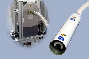 |
Bruker Transceiver Mouse Brain CryoProbe Coil Name: RF CP F2.5 500 1H M.BR1 Q S TR Part No. Z111531 |
Volume Coils:
 |
Bruker Volume Coil Coil Name: RF RES 500 1H 089/072 QUAD TO AD Description: Magnet south pole at the user end. Outer diameter is 89 mm, inner diameter is 72 mm. Part No. T11232V3 SNR:1624 (used as transceiver); 90 degree pulse: 14.0-14.9 db (depend on the surface coils combined) |
 |
Bruker PEX Volume Coil Coil Name: RF ARR 500 1H 089/072 8*1 TO AD Description: volume coil for Parallel Excitation Unit. Outer diameter is 89 mm, inner diameter is 72 mm. Part No. T20032V3 SNR:NONE |
 |
Bruker Mouse Body Array Volume Coil Coil Name: RF ARR 500 1H M.BODY 8*1 AD RO Description: Receive-only 1H mouse body 8 channel coil array, (with integrated preamplifiers, no tune /no match). Part No. T20033V3 SNR: 7413 (with volume coil T11232V3) |
 |
Bruker Rat Body Array Volume Coil Coil Name: RF ARR 500 1H R.BODY 8*1 AD RO Description: Receive-only 1H rat body 8 channel coil array, (with integrated preamplifiers, no tune /no match). Part No. T20035V3 SNR: NONE (with volume coil T11232V3) |
 |
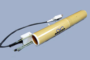
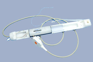
Bruker Rat Head/Mouse Body Volume Coil Coil Name: RF RES 500 1H 075/040 QSN TR Description: Quadrature 1H resonator for 500 MHz MR systems. Outer diameter is 75 mm, inner diameter is 40mm. Suitable as transceiver or as transmitter for cross coil experiments. It provides the correct circular polarization for the south pole of the magnet being at the patient end. This coil must be used in combination with the rat animal bed T12559. Part No. T11440V3 SNR: 6404; 90 degree pulse: 27.5 db |
 |
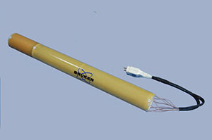
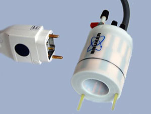
Bruker Mouse Head Volume Coil Coil Name: RF RES 500 1H 023 M.BR QSN TR Description: Circular Polarized MRI transceiver coil for 1H. Outer diameter 44 mm, inner diameter 23 mm. Distance from front plate to centre is 22 mm, i.e. the black line marked on the surface. This coil must be used in combination with the rat animal bed T12556. Part No. T12970V3 SNR: 12825 ; 90 degree pulse: 33.2 db |
Surface Coils:
 |
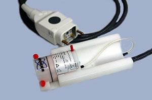
Bruker Mouse Brain Surface Coil Coil Name: RF SUC 500 1H M.BR QSN RO AD Description: 1H mouse brain receive-only surface coil, circular polarized with integrated combiner and preamplifier, no tune/ no match. It muse be used in combination with an actively detuned transmit-only resonator. Part No. T11657V3 SNR: 21707 (with T11232V3 volume coil ) |
 |
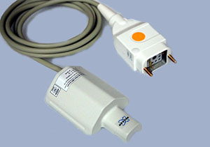
Bruker Rat Brain Surface Coil Coil Name: RF SUC 500 1H RATBR QSN RO AD Description: 1H rat brain receive-only surface coil, circular polarized with integrated combiner and preamplifier, no tune/ no match. It muse be used in combination with an actively detuned transmit-only resonator. Part No. T11425V3 SN:0029 SNR:7045 (with T11232V3 volume coil ) |
 |
Bruker Mouse Brain Surface Coil Array Coil Name: RF ARR 500 1H M.BR LIN RO AD Description: 1H mouse brain receive-only coil array (with integrated preamplifiers, no tune / no match). It must be used in combination with an actively detuned transmit-only resonator. Part No. T11766V3 SNR: 21163 (with T11232V3 volume coil ) |
 |
Bruker Rat Brain Surface Coil Array Coil Name: RF ARR 500 1H R.BR LIN RO AD Description: 1H mouse brain receive-only coil array (with integrated preamplifiers, no tune / no match). It must be used in combination with an actively detuned transmit-only resonator. Part No. T11473V3 SN: 0010 SNR:9757 (with T11232V3 volume coil ) |
 |
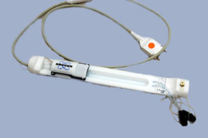
Bruker Mouse Cardiac Coil Array Coil Name: RF ARR 500 1H M.HRT RO AD Description: 1H mouse heat receive-only coil array 2*2 with integrated mouse bed and with integrated preamplifiers, no tune / no match. The coil is designed as RF coil attachment to the mouse animal bed. It must be used in combination with an actively detunned transmit-only resonator. Part No. T20029V3 SNR: 9515 (with T11232V3 volume coil ) |
Planar Surface Coils:
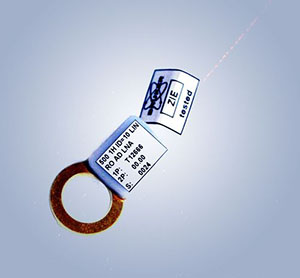 |
Bruker 10mm Planar Surface Coil Coil Name: RF SUC 500 1H ID=10 LIN RO AD LNA Description: Proton receive-only linear surface coil with diameter 10 mm. This coil is using simple active decoupling. It muse be used in combination with coil T20014V3. Part No. T12666 SN:0024 SNR: 25852 (with T11232V3 volume coil ) |
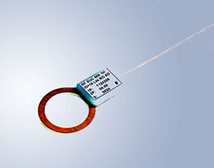 |
Bruker 15mm Planar Surface Coil Coil Name: RF SUC 500 1H ID=15 LIN RO AD Description: Proton receive-only linear surface coil with diameter 15 mm. This coil is using simple active decoupling. It muse be used in combination with coil T20014V3. Part No. T120209 SNR: 20547 (with T11232V3 volume coil ) |
 |
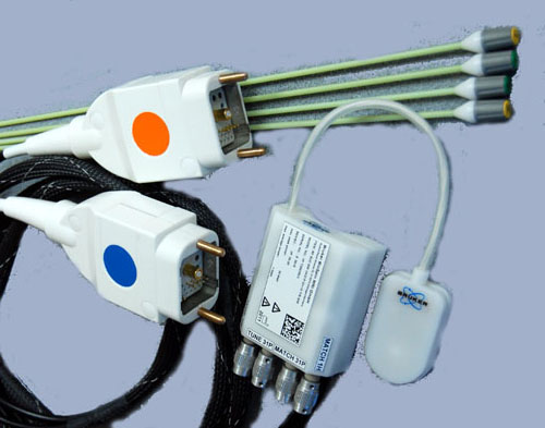
Bruker Low-noise Preamplifier Coil Name: RF SUC 500 1H LNA AV3 Description: The preamplifier is used for the surface coil T12666/T120209. Part No. T20014V3 |
Heteronuclear Coils:
 |
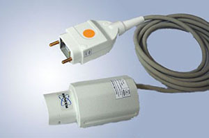
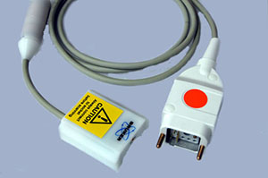
Bruker 1H/31P Transceiver Surface Coil Coil Name: RF SUF 500 1H/31P ID=10 T/R Description: heteronuclear, transmit/receive surface coils for 31P, equipped with cable, filters, T/R switch and preamplifier. Inner diameter 10 mm. Part No. T20039V3 SNR(1H): 8064;90 degree pulse (1H): 21.6 db |
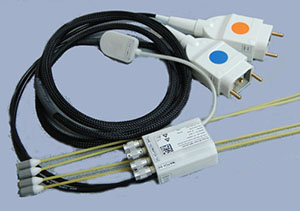 |
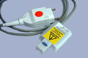
Bruker 1H/13C Transceiver Surface Coil Coil Name: RF SUF 500 1H/13C ID=10 T/R Description: heteronuclear, transmit/receive surface coils for 13C, equipped with cable, filters, T/R switch and preamplifier. Inner diameter 10 mm. Part No. T20036V3 SNR(1H): 5690; 90 degree pulse(1H): 19.6 db |
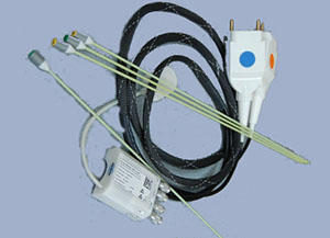 |
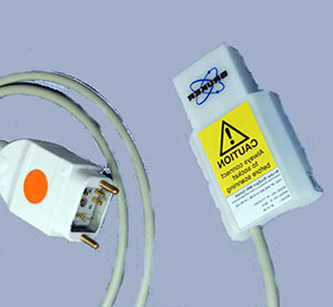
Bruker 1H/15N Transceiver Surface Coil Coil Name: RF SUF 500 1H/15N ID=10 T/R Description: heteronuclear, transmit/receive surface coils for 15N, equipped with cable, filters, T/R switch and preamplifier. Inner diameter 10 mm. Part No. T20037V3 SNR(1H):5569;90 degree pulse(1H):18.65 |
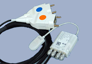 |
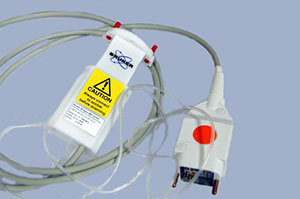
Bruker 1H/17O Transceiver Surface Coil Coil Name: RF SUF 500 1H/17O ID=10 T/R Description: heteronuclear, transmit/receive surface coils for 17O, equipped with cable, filters, T/R switch and preamplifier. Inner diameter 10 mm. Part No. T20038V3 SNR(1H):6418; 90 degree pulse(1H): 22.22 |
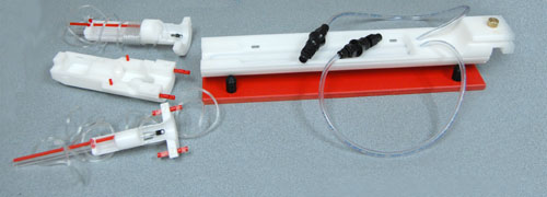 |
Autopac Mouse Bed |
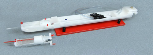 |
Autopac Rat Bed |
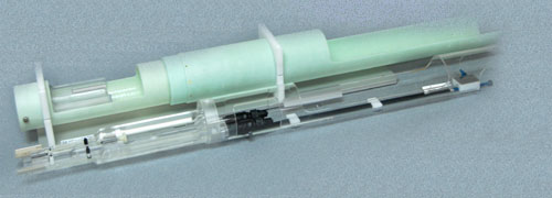 |
Animal Bed for Cyroprobe |
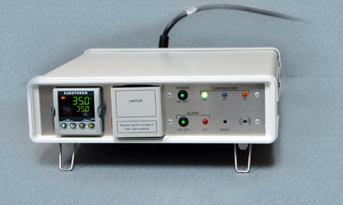 |
Temperature Control Unit of Cyroprobe This control unit allows monitoring of the temperature of the Animal / MRI CryoProbe interface and can keep it at any chosen temperature value. This unit has to be on during animal experiments, since the interface temperature will drop to zero Celsius without heater on. |
|---|---|
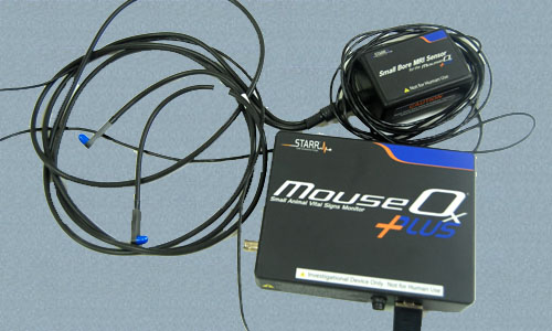 |
MouseOx Plus Oximeter The MouseOx Plus provides the following physiologic measurements: 1. Oxygen Saturation (SpO2); 2. Heart Rate; 3. Breath Rate; 4. Pulse Distention; 4. Breath Distention The Oximeter is available to all users. However, $5/hour will be charged to the users of the Oximeter to compensate the purchase cost and maintenance expenses. |
 |
Small Animal Monitoring and Gating System The monitoring system displays multiple waveforms and values, including ECG, Tempearture, Respiration and IBP. |
We offer the following manuals for the animal facility users, please contact xuj@kennedykrieger.org.
-
Paravision 5.0 Operation Manual
-
Paravision 5.0 Acquisition Manual
-
Paravision 5.0 Advanced Users Manual