By Taylor Gleason
For 26 years, the F.M. Kirby Research Center for Functional Brain Imaging at Kennedy Krieger Institute has been unlocking the mysteries of the brain.
Founded through a partnership with Johns Hopkins Medicine, the center was created to give scientists access to cutting-edge magnetic resonance imaging, or MRI, technology. What began with one scanner and a small team of experts has skyrocketed in scope to become one of the most important sites for brain research in the world.
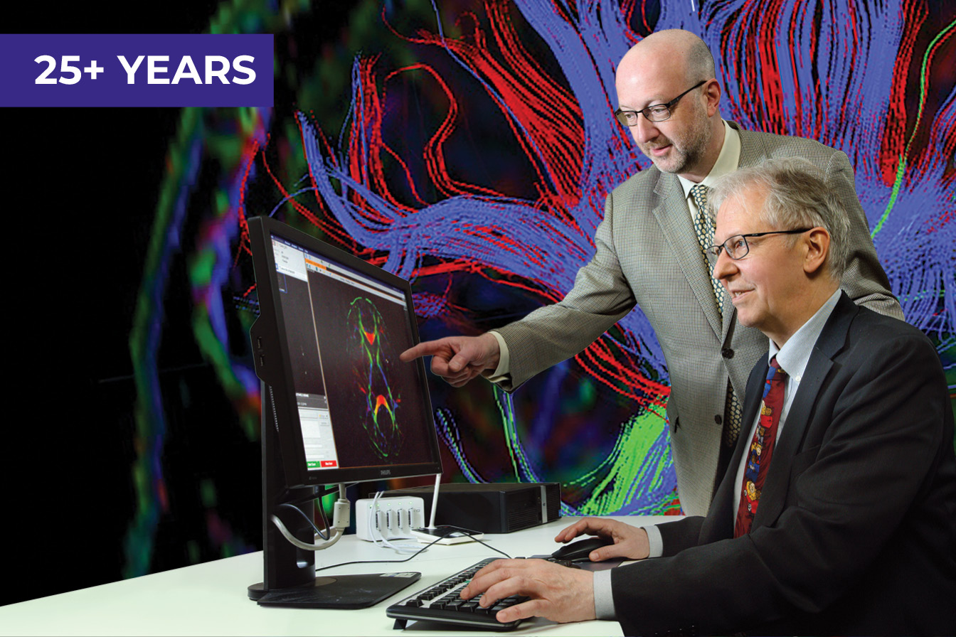
The center’s growth has been driven by major technological advances invented, in large part, by its own world-leading faculty and staff. Researchers have pioneered imaging techniques now used worldwide, from mapping brain connections to developing noninvasive ways to detect brain chemistry, tumor activity and even the brain’s communication with the gut. So far, its most important contributions to the medical field have been its complete map of human brain connections and its “MRI Atlas of Human White Matter,” the first full set of illustrations of connections in the living brain.
The heart of the Kirby Center is progress made possible by its dedicated faculty and support staff. The next 26 years truly hold limitless possibilities.” – Dr. Peter van Zijl
Today the center, which receives funding from the National Institute of Biomedical Imaging and Bioengineering, houses two 3T MRI scanners and a 7T fMRI scanner. The “T” is for “tesla,” a unit used to measure the force of a magnetic field, and the “f” stands for “functional,” because the scanner’s 60-ton magnet (housed in the Institute’s basement) is so strong it can track very small changes of activity in the brain by detecting changes in blood oxygenation.
Right now, the center’s faculty and staff are developing new methods for capturing multiple types of data in a single scanning session, to allow for personalized diagnosis and monitoring of treatment. It currently supports more than 150 active human research studies, among which is the HEALthy Brain and Cognitive Development Study, the largest long-term, in-depth look by MRI at early brain development in the U.S. In addition, the center is supporting over 50 preclinical projects—studies that test the effectiveness and safety of a drug or other treatment before it’s tested in people. The center also publishes books, papers and conference abstracts.
“The discoveries we’ve made and facilitated over the past 26 years are astounding—and life-changing,” says Dr. Peter van Zijl, the center’s director. “As we celebrate this milestone, our researchers are exploring the future of brain imaging. We are looking at everything from developing sugar-based imaging agents to harnessing AI to sharpen scan quality.” The center is just getting going—and its future holds ever-greater findings to come.
Did you know? The Kirby Center currently supports 150+ ongoing human studies and 50+ preclinical research projects from more than 10 departments at Kennedy Krieger and multiple schools at Johns Hopkins. The center’s techniques have been cited in tens of thousands of scientific papers.
Learn more about the F.M. Kirby Research Center for Functional Brain Imaging, which is a member of the MRI Resource for Physiologic, Metabolic and Anatomic Biomarkers, an interdisciplinary consortium combining Kennedy Krieger and Johns Hopkins Medicine facilities and expertise.