
The Kirby Center has three whole-body magnetic resonance imaging systems dedicated for research use.
Each MR scan room has been optimally designed for fMRI with attention to RF shielding, stimulus and response interface and environmental control:
- Optimum radio-frequency (RF) shielding: Soldered, monolithic copper shielding with air-sealed door is used to provide maximum protection from outside RF interference sources with long-term reliability. The rooms exceed the manufacturers specification for RF attentuation by as much as 30 dB.
- Two lab penetration panels with a variety of waveguides and filtered connectors provide the means to interface devices between the scan rooom and outside areas without compromising the RF shield. No RF-producing equipment is used in the scan rooms without proper shielding.
- 3T Scanners: The scan rooms are lined with layers of silicon steel sheet to reduce the influence of low frequency magnetic fields on imaging.
- 7T Scanner: A 400 metric tonne magnetic shield is used to limit the stray field generated by the magnet. The shield forms a six sided box encompassing most of the scan room varying in thickness from 8 to 16 inches.
- Air conditioning and power conditioning: Humidity and temperature in the scan rooms is controlled and monitored by dedicated equipment. 7T scanner's power is supplied by a dedicated uninterruptable power system (UPS) providing stable power condition and battery backup operation during brownouts or total power loss.
These systems are defined by the strength of their magnetic field, 3.0 Tesla or 7.0 Tesla. One Tesla is about 20,000 times stronger than the Earth's magnetic field. Each scanner has a thirty-two-channel receiver system and high-sensitivity multi-element receive-only head coils, allowing for parallel imaging, including sensitivity encoding (SENSE) for faster scanning.
3.0T MRI Systems
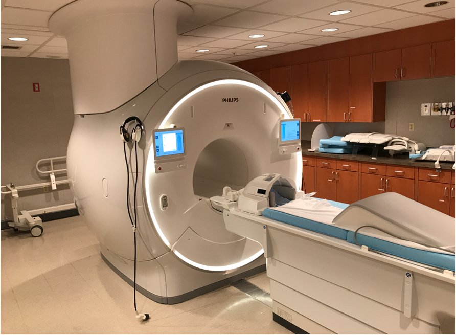
MR1 is a Philips dStream Achieva 3T TX; MR2 is a Philips dStream Ingenia Elition. These Philips 3 Tesla MRI scanners have many advanced and unique features to optimize physiologic, metabolic, anatomic and functional MRI studies, including:
- Short bores: 60 cm in diameter, 60 cm straight bore. Each bore flares in the front and back for a total magnet length of 179 cm cover-to-cover. A short bore provides a comfortable scanning environment, especially valuable for studies involving children, the elderly, and subjects with neurological and psychiatric disorders.
- Excellent field homogeneity: Even with the shorter bore, the main field is very homogeneous, which allows us to implement many modern imaging techniques.
- Modern imaging capabilities: Including echo planar imaging (EPI), fast spin echo (FSE), diffusion imaging with EPI and navigator echoes, single voxel MR spectroscopy (MRS), multi-slice spectroscopic imaging (MRSI), spectroscopic editing for neurotransmitters, compressed Sensing, multi-band (simultaneous multi-slice) acquisition.
- Gradients: On MR1, dual mode gradients run at 80 mT/m with a slew rate of 110 T/m/s, or at 40mT/m with a slew rate of 220 T/m/s. On MR2, the gradients were upgraded to 95 mT/m with a slew rate of 220 T/m/s in March 2021.
- Coils: These receive the magnetization signal.
- Both systems feature Philips dStream direct digital technology that samples the MR signal directly in the RF coil, transmitting the signal over fiber-optic cable to the reconstructor.
- Thirty-two channel head coil - Coil layout allows parallel imaging in AP, RL or head-foot direction.
- Twenty channel head and neck coil - excellent for neurovascular studies.
- Dual Flex-M coil - flexible surface coil, can be use in pairs.
- Small Extremity coil - 8 channel coil, 20 cm diameter.
7.0T MRI System
- This system was opened for use to our Center in April of 2009.
- 58 cm bore.
- Radiofrequency transmit channels allow heteronuclear and homonuclear spectroscopy with decoupling, interleaved with fast imaging.
- In November 2016, the system was upgraded with a second spectrometer that includes 8 independent transmit channels and an 8 channel head volume transmit coil. Using B1 calibration techniques, the 8 channels can be adjusted to provide more homogenous B1 excitations. The original 2 channel spectrometer is still available for backward compatibility.
- In January 2025, the system was updated to digital dSync architecture, Omega HP gradients with 40 mT/m and a slew rate of 200 T/m/s, compressed sense and simultaneous multislice (SMS) acquisition/reconstruction
- Shims: 3 first order (gradient), 5 second order and 7 third order shims plus 2 dynamically controlled shims
- Coils:
- Transmit/receive head coil - primarily used for QA.
- Thirty-two-channel receive head coil - (NovaMedical).
- Eight-channel receive spine coil (NovaMedical).
- Dual-tuned proton / 13C leg coil.
- Transmit/receive head coil - primarily used for QA.
- Volume TR head coil - normally used as the transmitter for 16 or 32 channel head coils (NovaMedical).
Thirty-two-channel receive head coil - (NovaMedical). - Eight-channel receive spine coil (NovaMedical).
- Dual-tuned proton / 13C leg coil.
This "mock scanner" allows researchers to do practice runs in order to acclimatize subjects to the sights and sounds of an MRI. It is especially useful for training children, the elderly, and persons with psychiatric disorders to perform an fMRI task and to reduce head motion during scanning. Our simulator includes:
- Model: A similarly-sized scanner cover by Philips Medical Systems.
- Surround sound audio system: This simulates the scanner sounds.
- Audio and video paradigm presentation system: The response buttons for these are identical to those used in the MRI scanner.
Dedicated Windows PCs are provided in each control room for fMRI stimulus presentation and response collection. They are connected to the scanners via custom triggering circuits. Macintoshes are available in both 3T control rooms.
Paradigm Presentation
Paradigm presentation software packages on our systems include:
- Psychology Software Tools E-Prime
- Neuro-Behavioral Systems Presentation
- MATLAB with the Psychophysics toolbox
Visual stimulation equipment:
- Video Projector - Epson 3 LCD projector, 1024x768 native resolution
- Computer connections
- DVD Recorder/Player connections
- Located in Equipment Room, projects into back end of scanner through 7 inch waveguide
- Remote control from Control Room
Audio stimulation equipment:
- Amplifier and equalizer
- Computer-generated sound
- DVD/CD player/recorder
- Philips in-room speakers
- Airtube headset with piezo-electric drivers
- Stax electrostatic ear speakers
- Sensimetrics piezo-electric headset
- Optoacoustics fiber-optic active-noise cancellation headset and microphone
Gauging Responses
Response monitoring equipment includes:
- fMRI-compatible fiber optic button boxes
- fMRI-compatible EEG equipment
- Eye-tracking of subjects inside the magnet: using infrared imaging
- Easily accessible interfaces: allowing investigator-owned computers to present paradigms to subjects
- Wave Guide Passthru connection
- Filtered DB25/DB9 connector
- Filtered BNC coaxial connector
fMRI Analysis Software Packages
- AFNI
- FSL
- SPM (running under MATLAB)
For software developed at the Kirby Center, please refer to our Software page.
We have several MR-compatible physiologic monitoring devices:
Philips devices for MR triggering and can be stored for fMRI correction
- Vector-electrocardiogram (VCG)
- Finger pulse
- Respiration pneuomgraph
Invivo Millennia 3500 MR compatible monitor
- Electrocardiogram (ECG)
- Peripheral pulse oximeter
- End tidal CO2
- Non-invasive blood pressure
- Invasive blood pressure
- Anesthetic inhalation gases
We have many computers, for storing data, and operating the scanner. Some specific details:
- Fast Gigabit Ethernet network
- File and compute server: Oracle M4000 server
-
- 32 cores
- 64GB of RAM
- Storage capability: 107 TB of RAID storage
- Compute Server: Penguin Computing, Inc. 21 node / 336 core Abu Dhabi Scyld IB Cluster
-
- Altus 2800i Master Node, Dual AMD Opteron 6320, 8 core, 2.8/3.3 GHz, 32 GB RAM, 1 TB RAID1 Boot Disk
- 2 x Altus 2840 Compute Nodes, 4 x Dual AMD Opteron 6320, 8 core, 2.8/3.3 GHz, 128 GB RAM / node, 1 TB scratch disk/ node,
- 3 x Altus 2840 Compute Nodes, 4 x Dual AMD Opteron 6320, 8 core, 2.8/3.3 GHz, 64 GB RAM / node, 1 TB scratch disk / node,
- Altus 2850GTi GPU Compute Node, Dual AMD Opteron 6320, 8 core, 2.8/3.3 GHz, 128 GB RAM, 1 TB scratch disk, 2 NVIDIA Tesla K20 GPUs
- All nodes interconnected through 40 GBps InfiniBand
- Scyld Clusterware HPC 6.0 cluster software
For several image analysis tools, please see the website for the Center for Imaging Science.