
Technology Research & Development #4: Algorithms for Functional and Anatomical Brain Analysis
Principal Investigators: Susumu Mori, PhD, and Michael Miller, PhD
The overall objective of TR&D4 is to develop cutting-edge image analysis technologies that can integrate various anatomical representations (multi-manifold) of the brain based on multi-modal MRI that includes not only multi-contrast anatomical MRI (T1, T2, diffusion, DTI, magnetization transfer, MT, susceptibility weighted and susceptibility tensor MRI, SWI and STI) but also functional MRI (fMRI) and MR spectroscopy (MRS) into a common anatomical framework. In addition, methods are developed to monitor time dependent anatomical change for growth (brain development) and neurodegeneration.
The multi-modal MR data will be co-registered within a common atlas space for:
- Multi-contrast quantitative analysis,
- Multi-manifold analysis for integration of anatomy and cortical function, and
- Cross-subject population-based analysis.
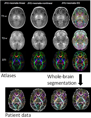
Image: Various types of human brain multi-modal atlases being developed for quantitative anatomical characterization of neonate brains. Depending on the applications, population-averaged (linear and non-linear versions) and single-subject atlases are being tested, which include T1-weighted, T2-weighted, and DTI contrasts. These atlases can be warped to patient images for comprehensive brain segmentation and automated quantitative reporting.