
Technology Research & Development #1: Quantitative MRS of the Brain at 3T and 7T
Principal Investigator: Peter Barker, DPhil
The metabolism of the central nervous system is of key interest for our driving biomedical projects in pediatric brain development, X-linked adrenoleukodystrophy, Alzheimer's disease, Multiple Sclerosis, Brain Tumors and Huntington's disease. In addition, several service projects (e.g. involving pediatric neurometabolic diseases such as Rett Syndrome, stroke, and HIV infection) involve the in vivo study of brain metabolite levels. Our overall goal is therefore to develop high-resolution MR Spectroscopic Imaging (MRSI) techniques for the study of brain and spine metabolism at the magnetic field strengths of 3 and 7 Tesla. This includes methods for the detection of neurotransmitters (such as GABA, glutamate, or N-acetyl aspartyl glutamate). Finally, for use in clinical practice and research, it is important that robust and easy to use data analysis tools exist for MRSI. We continue to develop quantitative analysis methods of MRS and MRSI data, and in particular (in collaboration with TR&D4) novel atlas-based techniques for statistical group analysis of MRSI data.
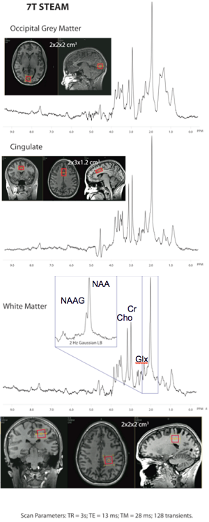
7T short TE STEAM spectra recorded using a Philips Achieva system with 32-channel receive head coil (Nova medical). 3 different brain regions (occipital gray matter, anterior cingulate gyrus, and posterior-frontal/parietal white matter were recorded. Scan time was 6min 24sec per region. Field honogeneity was optimized using high order shim coils. In the white matter voxel, the NAAG peak can be seen as a shoulder on the NAA resonance (inset).