
Technology Research & Development #2: Quantitative Multi-contrast MRI at 3T and 7T
Principal Investigators: Peter van Zijl, PhD, Hanzhang Lu, PhD
The discovery of tissue biomarkers that can provide early and more specific information regarding physiological or metabolic changes can be seen as the holy grail of clinical imaging. The availability of such biomarkers is one of the main issues that we are continuously being inquired about by our collaborative projects, which study such topics as autism spectum disorder (ASD), pediatric brain development, Attention Deficit Hyperactivity Disorder (ADHD), Alzheimer’s Disease (AD), Multiple Sclerosis (MS), Huntington’s Disease (HD), Schizophrenia (SCZ), and brain tumors. Our overall goal is therefore to develop MRI methodologies at 3T and 7T that can provide the quantitative biomarkers needed by our CPs. Examples of quantitative parameters are magnetization transfer constants (both exchange transfer and cross-relaxation based), magnetic susceptibility constants derived from frequency maps, and cerebral blood volumes and the metabolic rate of oxygen metabolism.
Contents:
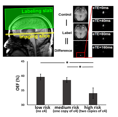
The brain represents 2% of the body weight but consumes 20% of the total energy. Therefore, the rate of oxygenextraction and consumption is a sensitive index of brain function and tissue viability. Non-invasive imaging of brain oxygenation and metabolism can provide an early marker in brain diseases such as Alzheimer’s disease, before clinical symptom can be detected. Using a novel pulse sequence, TRUST MRI, the brain’s oxygen extraction fraction (OEF) was measured in three groups of cognitively normal elderly individuals, but with different levels of genetic risk (Apo E) for Alzheimer’s. It was observed that, even in the absence of any clinical symptoms or cognitive deficits, OEF measured with TRUST MRI can already detect a significant reduction in brain oxygen extraction in high-risk individuals. Aside from Alzheimer’s disease, brain metabolic techniques have also found applications in Multiple Sclerosis, aging, neonates, anorexia, glucose transporter deficiency, sickle cell anemia, and cocaine addiction.
Data from:
Hanzhang Lu, Zixuan Lin, Peter van Zijl, and Marilyn Albert
Department of Radiology, Johns Hopkins University School of Medicine
F.M. Kirby Research Center for Functional Brain Imaging at Kennedy Krieger Institute
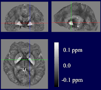
These high-resolution images (1 microliter volume elements) depict the magnetic susceptibility properties of the brain. The contrast in these images reflects "how magnetic" the parts of the brain are, relative to water. In the deep gray matter nuclei, where iron content is high, hyperintensity is clear, while the white matter bundles appear dark. Iron content in gray matter increases with age. Increased brain iron concentration has also been implicated in neurodegenerative diseases such as Alzheimer's Disease, Parkinson's Disease and Huntington's Disease. This type of imaging may provide insight into developing methods for early detection of such diseases.
Images from:
Xu Li, Issel Anne Lim, Craig Jones, Peter van Zijl
F.M. Kirby Research Center for Functional Brain Imaging at Kennedy Krieger Institute
Department of Radiology, Johns Hopkins University School of Medicine, Baltimore, MD
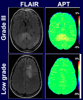
Low-grade and high-grade brain tumors can be compared with conventional clinical MRI (FLAIR) images and with a novel protein-based approach called Amide-Proton Transfer (APT) MRI. Low- and high-grade images appear similar on conventional MRI, but can be distinguished using this new type of molecular imaging, which exploits endogenous changes in protein content. APT images also have the potential to monitor the effects of tumor therapy.
Image provided by:
Jinyuan Zhou, Jaishri Blakeley, John Laterra and Peter van Zijl
F.M. Kirby Research Center for Functional Brain Imaging at Kennedy Krieger Institute
Department of Radiology, Johns Hopkins University School of Medicine, Baltimore, MD