
Development of techniques to measure basal physiological parameters
Next-generation ASL techniques – Velocity-selective ASL and MR Fingerprinting ASL
Cerebral perfusion imaging plays a pivotal role in many brain diseases. Arterial Spin Labeling (ASL) MRI techniques, such as pseudo-continuous ASL (PCASL), allow for non-contrast evaluation of CBF by magnetically labeling water in the arterial blood as an endogenous tracer. Although PCASL has been widely used in clinical applications, this technique is susceptible to variations in bolus arrival time (BAT). This confounding factor is particularly problematic in older individuals or in patients with vascular conditions such as vessel narrowing. We are developing ASL techniques that are immune to variations in BAT, thereby providing more reliable estimation of CBF.
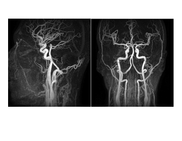 (i) Velocity-selective ASL (VS-ASL) aims to label arterial blood immediately adjacent to the imaging voxel via a blood velocity-selection sequence module, thus rendering the signal insensitive to BAT and also alleviating signal loss (i.e. T1 recovery) due to transit delay. Techniques include inversion velocity-selective ASL, which can further enhance the sensitivity of the technique (compared to saturation velocity–selective methods). Additional designs will include Fourier Transform (FT)-VS pulse trains that have much improved immunity to magnetic field inhomogeneity, which is achieved by inserting paired and phase-cycled refocusing pulses within each velocity-encoding step. The image on the right shows an example of VS-based MR angiogram.
(ii) MR Fingerprinting ASL allows simultaneous estimation CBF, BAT, tissue T1, and cerebral blood volume (CBV), among other parameters, i.e. multi-parametric estimation of brain hemodynamics, in a single scan of 4 minutes, based on the principle of MR Fingerprinting (MRF).
Oxygen extraction fraction (OEF) techniques:
Cerebral oxygen extraction fraction (OEF) is a key physiological index of the homeostasis of oxygen demand and supply in the human brain. Quantitative assessment of OEF is of potential clinical value for diagnosis and treatment monitoring of neurological conditions. Positron Emission Tomography (PET) with 15O-labeled tracers has been used extensively for OEF determination, but its use overall is still limited due to the need for an on-site cyclotron, arterial sampling, and the exposure to ionizing radiation. Techniques measuring cerebral OEF without any exogenous agent on standard MRI scanners within just a few minutes can enable the routine assessment of OEF in human patients.
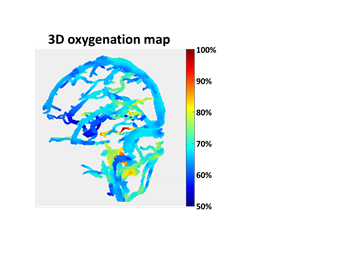 (i) T2-based OEF techniques: We have previously developed a T2-Relaxation-Under-Spin-Tagging (TRUST) MRI sequence that measures OEF globally, which has found broad clinical applications and has been requested and used by more than 40 laboratories world-wide. Current pulse sequence development work has the goal of obtaining 3D, whole-brain mapping of OEF in <5 minutes. To make this feasible, we will develop accelerated OEF acquisition schemes by incorporating turbo-field-echo (TFE), compressed sensing (CS), variable-density sampling, and arterial nulling methods. The image on the right shows an example of a 3D oxygenation map.
(ii) Quantitative susceptibility mapping (QSM) based OEF technique: We are also developing QSM-based techniques to measure blood oxygenation in veins, for diseases with atypical hemoglobin such as sickle cell disease where the conventional T2-oxygenation calibration plot may not be valid. Since blood magnetic susceptibility has a simply linear dependence on the blood oxygenation fraction and Hb concentration, it is possible to incorporate the sickle-Hb information into the estimation of blood oxygenation, making QSM based OEF measurement potentially more reliable in SCD patients.
Blood-brain barrier (BBB) permeability techniques
Microvasculature of the brain possesses a vascular barrier, called the blood brain barrier (BBB). An intact BBB is critical in protecting the brain from infection and inflammation. Disruption of the BBB has been associated with many pathological conditions. The ability to transiently maneuver the BBB permeability has also been exploited for efficient drug delivery in brain tumors, lysosomal storage disease and Parkinson’s disease. Therefore, a technique to report on BBB permeability in humans will have a tremendous potential in clinical applications. Previous research on BBB permeability was based largely on the metallic contrast-agent Gd-DTPA. However, Gd-DTPA is 30 times larger than water molecule and is only sensitive to major breakdowns of BBB at advanced stage of a disease. In order to detect early injury to BBB, techniques to evaluate its permeability to smaller molecules is desirable.
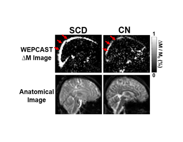 Water-extraction-with-phase-contrast-arterial-spin-tagging (WEPCAST) MRI features a phase-contrast velocity-encoding module in the ASL acquisition, which is specifically designed to measure ASL signal in venous vessels without partial volume effects of tissue perfusion signal. This allows probing water exchange rate, and thus BBB permeability to water non-invasively. The technique is first being developed for global BBB assessment through measurements in the superior sagittal sinus. The figure on the right shows WEPCAST images from a pediatric patient with sickle cell disease (SCD) and an age-matched healthy child. Further developments are planned to make the approach applicable for smaller veins (e.g. internal cerebral veins, the vein of Galen) that drain specific brain regions.