
CVR mapping without needing gas-inhalation.
Cerebrovascular Reactivity (CVR), the ability of the blood vessels to dilate or constrict, represents an important domain of vascular function and, compared to baseline vascular parameters, is expected to be more specific in reflecting vascular health. Current CVR techniques are based on CO2 gas inhalation that manipulate CO2 concentration in the blood. CVR has received increasing attention in clinical neuroscience due to its sensitivity to vascular abnormalities. However, its wide applications have been hampered by the procedural complexity of needing to delivery CO2 gas to the participant inside the MRI scanner. Therefore, we are developing CVR mapping techniques without gas-inhalation.
(i) Resting-state CVR mapping: We will develop an optimized strategy for rs-CVR mapping. Rather than manipulating the EtCO2 level by hypercapnia breathing, this approach exploits the natural variations in breathing pattern and depth of the participant over time, which results in fluctuations in end-tidal CO2. This method will allow for a broader application of CVR in routine clinical practice. Given the wide availability of resting-state fMRI data, it will also allow investigators to use our method to retrospectively estimate CVR.
(ii) CVR mapping method with intermittent breath modulation: Since a potential limitation of resting-state based CVR mapping is that the SNR may not be good in some participants, the intermittent breath modulation enhances the variations in breathing pattern without adding much burden to the participants. Therefore, this method combines the advantages of both resting-state CVR (e.g. comfortable to the participants, easy to conduct) and gas-based CVR (e.g. good sensitivity), and will further broaden the scope of applications of CVR in clinical settings.
Arterial pulsatility and vascular compliance mapping
Vascular compliance (VC) denotes the ability of a vessel to distend passively in response to an increase in blood pressure. A normal VC in major arterial vessels is important in attenuating pulsatile blood flow in large arteries into continuous flow in capillaries, thereby protecting the microvascular bed and parenchyma from injuries due to flow pulsatility. A diminishment in vascular compliance, i.e. vessel stiffness, has been suggested to be a risk factor for a number of diseases, and may result in brain pathology. Measurement of vascular pulsatility/compliance is a new direction in the brain MRI field. Although single-slice, 2D MRI imaging methods have been reported, a high-resolution 3D method for vascular pulsatility and compliance mapping is desirable.
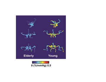 We are developing an MRI technique that allows quantitative mapping of VC in major intracranial arteries. This technique uses PCASL labeling, radial acquisition, and compressed sensing to measure arterial vessel volume in a spatially and cardiac-phase resolved manner, yielding a vascular pulsatility measure. This measure, when combined with pulse pressure, then provides an estimation of VC. The image on the right illustrates VC maps in a younger (30 years; male) and an older (75 years, female) participant.