

Nebel, M. B., Joel, S. E., Muschelli, J., Barber, A. D., Caffo, B. S., Pekar, J. J., & Mostofsky, S. H. (2014). Disruption of functional organization within the primary motor cortex in children with autism. Human Brain Mapping, 35, 567-580. doi:10.1002/hbm.22188
Anatomic Imaging
The Neuroimaging Core interacts closely with the F.M. Kirby Research Center for Functional Brain Imaging at Kennedy Krieger, which houses two Philips 3 Tesla MRI scanners and a Philips 7 Tesla scanner dedicated exclusively to research. We have developed and now can apply detailed and automated parcellation protocols of cortical and subcortical structures, high-dimensional registration methods for regional and whole brain parcellation, and sophisticated approaches of surface deformation mapping for highly localized regional examination of shape and thickness. In addition, we have developed a parcellation protocol for the cerebellum. In collaboration with the Center for Imaging Science in the School of Engineering at Johns Hopkins (PI: Dr. Michael Miller), we also have developed high dimensional registration methods to improve the delineation and definition of multiple brain structures/regions that often play roles in anomalous brain development and developmental disability.
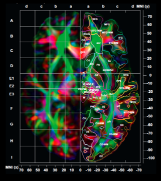
One of the challenges that almost inevitably confronts investigators who use neuroimaging tools is the overwhelming volume of data generated during image acquisition. In addition to supporting the range of “standard” procedures for analysis and interpretation, Dr. Susumu Mori and his colleagues have developed and now offer an anatomical framework in which the entire brain is segmented to about 200 structures, providing a major reduction in the size of informative data sets. These segmentation criteria are shared for all imaging modalities, so that the information can be integrated.
More generally, those who use neuroimaging investigative tools need access to a properly designed and executed imaging pipeline. One of the critical activities of the Neuroimaging Core is guiding its users so that they can accomplish this goal. Finally, the core provides a computational and data storage infrastructure, and through support from the Brain Science Institute at Johns Hopkins, the core provides free image analysis to generate preliminary data.
Small Animal Imaging
In addition to facilities for supporting MRI protocols in “human subjects,” Dr Susumu Mori supervises the Kirby Center animal imaging facilities. These resources are used by IDDRC-supported investigators in studies of CNS development and animal models of developmental disorder, employing 11.7T and 17.6T MRI for research needing especially high resolution.
Functional MRI (fMRI)
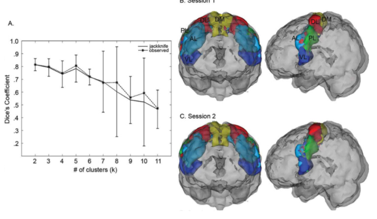
Functional MRI has become a widely applied technique. It allows for the localization of neural activity related to an extremely broad variety of cognitive and sensorimotor tasks. Thus, fMRI allows an investigator to understand the link between brain structure and function. In addition to supporting work related to “standard” (task-activation) fMRI, we also guide Neuroimaging Core users through the technical intricacies of applying the more recently developed class of techniques known as resting state functional connectivity MRI (rsfc-MRI).
Proton Magnetic Resonance Spectroscopy
Proton magnetic resonance spectroscopy (1H-MRS) enables non-invasive detection of neurochemicals at concentrations in the high micromolar to millimolar range. MRS techniques have thus made possible detection and reliable quantitation of neurochemicals that are of interest in studies of neurodevelopmental or psychiatric disorders of childhood and adolescence. In addition, this aspect of the core provides potential treatment targets. Given the increased MRS signal resolution afforded by scanning at 7T, and the wide range of potential applications, Dr. Alena Horska, one of our core investigators, has spearheaded efforts to facilitate the scanning of children in the 7T scanner without sedation.
Trans-cranial Magnetic Stimulation (TMS)
The various MRI-based methods supported by the core represent powerful tools for studying brain structures and functions, and TMS provides additional capabilities for IDDRC researchers. Here, localized high-field magnetic stimulation can be used either to enhance or inhibit brain activity associated with specific cognitive functions. Although considerable advances have been made independently in TMS, few facilities are available for exploring the potential of combining these methods for investigating connectivity in the human brain. We are using, and continuing to develop, techniques in which TMS and MRI are used together.
Educational Support
Education of users is essential for execution of proper image analysis. The monthly education and tutorial by Dr. Mori and his staff members enrolls approximately 150 users each year. This tutorial introduces the participants to both the principles of different types of anatomic imaging (including diffusion tensor imaging) and the “nut and bolts” of applying analysis software packages. Once the users take the two-day tutorial, they are entitled and encouraged to participate in face-to-face education sessions that focus on challenges related to their own MRI data.